
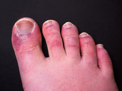
COVID-19 infection has been associated with a variety of rashes, some of which resemble frostbite and have been referred to as “COVID toes”. Frostbite is an ischemic phenomenon, caused by lack of blood supply to the digits for a prolonged period of time. Whether these rashes are in fact ischemic remains to be determined, due to the paucity of data from biopsy and other invasive assessments. COVID-19 patients have also been observed to deteriorate abruptly approximately one week after the onset of symptoms due to severe, widespread clotting within the blood vessels. Any relationship between ischemic-appearing skin findings and this ischemic cause of multi-organ failure remains unknown. Yet another complication of COVID-19 infection that could result in rashes as well as widespread ischemia is vasculitis (inflammation of the blood vessels). Due to the invasive nature of biopsies and the rapidity with which death occurs in fulminant cases, hyperspectral imaging may provide a more effective way to assess rashes for ischemia and help differentiate dysfunctional clotting from vasculitis, which may benefit from different treatments.
The programme seeks to develop a non-invasive and rapid method to assess skin findings for ischemia; categorise rashes based on spectral signatures; and correlate different rash subtypes with clinical outcomes.
Principal investigator Dr Raja Srinivasan, Clinical Assistant Professor at the University of Maryland School of Medicine, said, “Hyperspectral imaging is a wonderful tool for analysing COVID-19 rashes. It combines the power of spectroscopy with digital imaging to identify subtle wavelength differences that are not observable by the human eye. The high spectral and spatial resolution achievable with the HinaLea imager will hopefully provide a calibrated and reproducible method for assessing rashes across a wide range of skin pigmentation. The identification of spectral signatures that correlate with different pathologic mechanisms would further demonstrate the value of hyperspectral imaging as a tool for virtual biopsy, providing key information for medical decision-making without the invasive and time-consuming nature of traditional tissue biopsy.”
The project started in June 2020 at two designated hospitals in Maryland and New York. Data will be collected from up to 200 critically-ill COVID-19 patients.
“Imager portability and ease of use is extremely important for this programme because the data will be collected in a busy, time- and space-constrained clinical environment”, said Alex Fong, Vice President of Engineering at HinaLea. “In a truly transformative development, HinaLea has developed the only hand-held hyperspectral imager in the world that is capable of contact measurement. For the user, it is as easy as point-and-click.”

 located just 3 m from the exit nozzle of the engine. The testing facility is at the Instituto Nacional de Tecnica Aeroespacial (INTA) in Madrid. Credit: Gordon Humphries, University of Strathclyde")


 nano-DESI mass spectrometry imaging of liver tissue from orally dosed rat (Animal 3). a) optical image of a blood vessel within liver tissue. b) Composite ion image of charge-reduced haeme-bound α-globin (7+ and 6+ charge states; m/z 2259.9 and m/z 2636.3 respectively, red) and the charged-reduced [FABP+bezafibrate] complex (7+ and 6+ charge states; m/z 2097.5 and m/z 2446.9 respectively, blue). c) Ion image composed from charge-reduced haeme-bound α-globin (7+ and 6+ charge states) showing abundance in blood vessels. d) Ion image composed from charge-reduced [FABP+bezafibrate] complex (7+ and 6+ charge states) showing abundance in bulk tissue and absence in the blood vessel. Reproduced from https://doi.org/10.1002/ange.202202075 under a CC BY licence.")
, Prof. Dr. Wolfgang Kiefer with the historical portrait of C.V. Raman, Dr. Joachim Koenen (WITec Managing Director).")