

Mass spectrometry imaging (MSI) provides highly precise information on the spatial distribution of substances in many areas. Researchers at the University of Bayreuth have succeeded in making visible an additive in dairy products and a production-related contamination in baked goods. Special ingredients that influence food quality can be detected in fruit, vegetables and meat products. The study, which was conducted in cooperation with the Bavarian Health and Food Safety Authority (LGL), shows the great potential of this method, not least in terms of consumer protection.
Natamycin in cheese
To protect cheese wheels or smoked sausages from mould infestation, the surfaces are often treated with the fungicide natamycin. An EU regulation sets a limit of one milligram per square decimetre for this and also stipulates that natamycin must not penetrate deeper than 5 mm into a treated cheese wheel. However, this penetration depth cannot be described in detail using the food analysis methods commonly in use. However, the Bayreuth research team led by Prof. Dr Andreas Römpp has been able to use MSI to show for the first time where and in what quantities the fungicide occurs in different types of Gouda. The penetration of the natamycin molecules can be tracked from the rind to the inside of the cheese wheel. The scientists collaborated with the LGL in these investigations. Based on the results obtained, they have developed methodological standards for the identification of natamycin in cheese”, says Prof. Römpp, who is the Chair of Bioanalytic Sciences and Food Analysis at the University of Bayreuth.
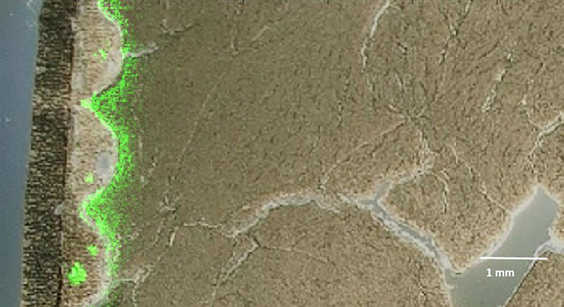
The fungicide natamycin (green) penetrates inward from the coating (left) in some locations. Credit: © Julia Kokesch-Himmelreich
Acrylamide in gingerbread
An EU regulation also sets limits for the presence of acrylamide in food. It is a cancer-promoting substance that is formed from sugar and asparagine at low humidity and temperatures above 120°C. A method developed in Bayreuth, Germany, based on MSI, visualises acrylamide distribution in traditional German gingerbread. “To do this, we had to cool the gingerbread samples to less than ‒60°C and then use an electric microsaw to produce gingerbread slices of 2 mm thickness. This was the only way we could detect very small amounts of acrylamide”, reports Prof. Römpp.
The new study also shows that MSI is equally suitable for analyses of processed meat products. In veal sausages, water-soluble and fat-soluble components become visible, so that low-fat and high-fat regions can be clearly distinguished. Likewise, it becomes visible where substances of plant origin are found that come from admixed herbs. “However, MS imaging not only enables the localisation of ingredients in meat products, but also helps, for example, in investigations of ‘sticky meat’ or so-called hydrolysate additives, which are supposed to feign higher quality when they are not declared on the packaging. It could therefore be useful in detecting consumer deception in meat products and better protect consumers in this respect as well”, says Prof. Römpp.
Kiwifruit and carrots
The application potential in the field of fruits and vegetables is demonstrated by studies on kiwifruit and carrots. The “mini kiwi” (Actinidia arguta) is not only sweet, but also has numerous health-promoting bioactive ingredients. Using sample slices that were only a few hundredths of a millimetre thick and cooled down to a temperature of ‒40°C, the Bayreuth bioanalysts visualised the distribution of several substances in the skin and flesh: sugar molecules (disaccharides), antioxidant polyphenol and a fat (lipid) characteristic of kiwis. In carrots, in turn, molecules of beta-carotene, a precursor of vitamin A, were detected. In addition, it was also possible to identify the spatial distribution and typical molecular structures of different dyes (anthocyanins) that give carrots an orange, yellow or violet coloration.
“Our study makes it clear that MS imaging is a valuable addition to already established food analysis methods: it offers new insights into the spatial distribution and relative proportions of ingredients. It has the great advantage that the molecules of the ingredients do not have to be labelled with dyes or other labelling methods. At the University of Bayreuth—within the newly established Faculty VII of Life Sciences: Food, Nutrition and Health—we will continue to work in the future on refining the analytical capabilities of imaging mass spectrometry, combining it with other food analysis tools, and applying it to ingredients not previously studied. In this way, we at the University of Bayreuth can make important contributions to consumer protection”, says Prof. Römpp.


 cells in treated tumour cells")


 generate an RGB video image, the newly developed laparoscope (left) uses a multispectral camera. This also makes it possible to visualise functional properties of the tissue. Source: Leonardo Ayala / DKFZ")