
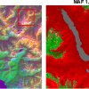
 Schematic of scanning Raman picoscopy (SRP). When a laser beam is focused into the nanocavity between the atomically sharp tip and substrate, a very strong and highly localised plasmonic field will be generated, dramatically enhancing the Raman scattering signals from the local chemical groups in a single molecule right underneath the tip. (b) Merged SRP image by overlaying four typical Raman imaging patterns shown on the right insets for four different vibrational modes. (c) Artistic view of the Mg-porphine molecule showing how four kinds of chemical groups (coloured “Legos”) are assembled into a complete molecular structure. ©Science China Press")
Precise determination of the chemical structure of a molecule is of vital importance to any molecular related field and is the key to a deep understanding of its chemical, physical and biological functions. Scanning tunnelling microscopy and atomic force microscopy have outstanding abilities to image molecular skeletons in real space, but these techniques usually lack chemical information necessary to accurately determine molecular structures. Raman scattering spectra contain abundant structural information of molecular vibrations. Different molecules and chemical groups exhibit distinct spectral features in Raman spectra. Therefore, the above deficiency can, in principle, be overcome by a combination of scanning probe microscopy with Raman spectroscopy, as demonstrated by tip-enhanced Raman spectroscopy (TERS), which opens up opportunities to determine the chemical structure of a single molecule.
In 2013, a research group led by Zhenchao Dong and Jianguo Hou at the University of Science and Technology of China (USTC) demonstrated sub-nanometre resolved single-molecule Raman mapping for the first time (https://doi.org/10.1038/nature12151), reducing the spatial resolution with chemical identification capability down to ~5 Å. Since then, researchers around the world continued to develop such single-molecule Raman imaging techniques to explore what is the ultimate limit of the spatial resolution and how this technique can be best utilised.
Recently, the USTC group published a research paper in National Science Review (NSR) entitled “Visually Constructing the Chemical Structure of a Single Molecule by Scanning Raman Picoscopy”, pushing the spatial resolution to a new limit and proposing an important new application for the state-of-art technique. In this work, by developing a cryogenic ultrahigh-vacuum TERS system at liquid-helium temperatures and fine-tuning the highly localised plasmon field at the sharp tip apex, they further drive the spatial resolution down to 1.5 Å on the single-chemical-bond level, which enables them to achieve full spatial mapping of various intrinsic vibrational modes of a molecule and discover distinctive interference effects in symmetric and anti-symmetric vibrational modes. More importantly, based on the Ångström-level resolution achieved and the new physical effect discovered, by combining with Raman fingerprint database of chemical groups, they further propose a new methodology, coined as Scanning Raman Picoscopy (SRP), to visually construct the chemical structure of a single molecule; see (a).
By applying the SRP methodology to a single magnesium porphyrin model molecule, the researchers at USTC obtained a set of real-space imaging patterns for different Raman peaks, and found that these patterns show different spatial distributions for different vibrational modes. Taking the typical C–H bond stretching vibration on the pyrrole ring as an example, for the anti-symmetric vibration (3072 cm–1) of two C–H bonds, the phase relation of their local polarisation responses is opposite. When the tip is located right above the centre between two bonds, the contributions from both bonds to the Raman signals cancel out, giving rise to the “eight-spot” feature in the Raman map for the whole molecule, with the best spatial resolution down to 1.5 Å. These “eight spots” have good spatial correspondence with the eight C–H bonds on the four pyrrole rings of a magnesium porphyrin molecule, indicating that the detection sensitivity and spatial resolution have reached the single-chemical-bond level. Raman imaging patterns of other vibrational peaks also show good correspondence to relevant chemical groups in terms of characteristic peak positions and spatial distributions [as shown in (b) and (c)]. The correspondence provided by the simultaneous spatially and energy-resolved Raman imaging allows them to correlate local vibrations with constituent chemical groups and to visually assemble various chemical groups in a “Lego-like” manner into a whole molecule, thus realising the construction of the chemical structure of a molecule.
Scanning Raman picoscopy (SRP) is the first optical microscopy that has the ability to visualise the vibrational modes of a molecule and to directly construct the structure of a molecule in real space. The protocol established in this proof-of-principle demonstration can be generalised to identify other molecular systems, and can become a more powerful tool with the aid of imaging recognition and machine learning techniques.

 located just 3 m from the exit nozzle of the engine. The testing facility is at the Instituto Nacional de Tecnica Aeroespacial (INTA) in Madrid. Credit: Gordon Humphries, University of Strathclyde")


 nano-DESI mass spectrometry imaging of liver tissue from orally dosed rat (Animal 3). a) optical image of a blood vessel within liver tissue. b) Composite ion image of charge-reduced haeme-bound α-globin (7+ and 6+ charge states; m/z 2259.9 and m/z 2636.3 respectively, red) and the charged-reduced [FABP+bezafibrate] complex (7+ and 6+ charge states; m/z 2097.5 and m/z 2446.9 respectively, blue). c) Ion image composed from charge-reduced haeme-bound α-globin (7+ and 6+ charge states) showing abundance in blood vessels. d) Ion image composed from charge-reduced [FABP+bezafibrate] complex (7+ and 6+ charge states) showing abundance in bulk tissue and absence in the blood vessel. Reproduced from https://doi.org/10.1002/ange.202202075 under a CC BY licence.")
, Prof. Dr. Wolfgang Kiefer with the historical portrait of C.V. Raman, Dr. Joachim Koenen (WITec Managing Director).")