Frank Neubrech and Annemarie Pucci
Kirchhoff Institute for Physics of the Heidelberg University, Im Neuenheimer Feld 227, 69120 Heidelberg, Germany
Introduction
Infrared (IR) spectroscopy of vibrational excitations is an extremely powerful technique because it combines sensitive chemical analysis with an almost non-destructive probe. High spectral resolution goes along with a dynamic range of at least four orders of magnitude in photometric sensitivity. Furthermore, modern spectrometers allow very fast measurements and, for example, reasonable monolayer spectra can be acquired today within a second. In such a measurement, typically, the focal spot amounts to 1 cm2, which means that, for a monolayer about 1015 molecules contribute to the measured signal. In a sensing application in which a lower number of molecules is present, IR spectroscopy is hampered by the rather tiny vibrational absorption cross section which is much smaller than the IR wavelength squared. This situation can be improved by local field enhancement.
Surface-enhanced infrared absorption
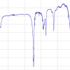
It is well known that a good metallic substrate in an IR reflection–absorption spectroscopy (IRRAS, with p-polarised light under grazing incidence) measurement enhances vibrational signals up to two orders of magnitude compared with normal transmittance or reflectance on insulators. In 1980, Hartstein et al.1 found that the IR absorption of molecules on metal films can be enhanced even more. The enhancement factor can reach the order of 1000 if the molecules are adsorbed onto Au or Ag island films.2,3,4 This effect is called surface-enhanced infrared absorption (SEIRA). The strength of the electro-magnetic enhancement strongly depends on the IR excitation spectrum of the metal islands (in fact it is a plasmonic excitation) and, thus, on metal–film morphology. Maximum plasmonic enhancement is found for metal–island layers near percolation.5,6 Model calculations based on effective dielectric functions (for example, on the Bruggeman model) are able to reasonably describe the enhanced vibrational spectra for very small (i.e. not scattering) metal islands and the change in adsorbate line-shape with metal-filling factor.5 Vibrational lines with the strongest enhancement appear with an asymmetric shape, see Figure 1. The asymmetry is considered to be the consequence of a Fano-type effect from the dipolar interaction of the vibrational excitation with the very broad plasmonic resonance of the metal particle ensemble. Near-field enhancement is an important issue for the coupling. The plasmonic excitation is localised at the surface region of the metal particles and near-field enhancement benefits from low electronic damping, especially in Cu, Ag and Au in the IR and in the case of larger crystalline grains. For the electric field parallel to the layer, near-field enhancement is strongest in narrow gaps between islands and can be estimated from the ratio of particle width to gap width.
 on an Cu-island film on KBr(001); left, deformation modes; right, C–H stretching modes including a tiny surface mode at 2958 cm–1. Compared to ethane ice, IR signals are, on average, enhanced by about 100 times. Reference is the bare island layer. It was deposited under ultra-high vacuum conditions at room temperature and corresponds to 9 nm average thickness. The inset shows an atomic-force microscopy image of a Cu-island film (500 nm × 500 nm) with similar thickness on KBr(001). The metal particle layer has a nearly frequency independent mid-IR transmittance of about 31%.")
Surface-enhanced infrared scattering
Considerably higher signal enhancement can be achieved with micrometre long plasmonic nanowires, called nanoantennas, under resonance conditions.7 Such big objects not only absorb but also scatter IR light. With IR micro-spectroscopy the far-field extinction spectrum of one particle can be measured, for example in normal transmittance with a transparent substrate. Usually, the substrate transmittance serves as reference. For plasmonic nanowires with diameter (typically 50 nm to 100 nm) much smaller than the length, the resonance photon wavelength in the mid-IR region is almost linearly proportional to the nanowire length L, since resonances correspond to standing waves in the surface region.8 Deviation from ideal antenna behaviour is related to the finite penetration of IR light into metal. By changing L, nanowire resonances can be tuned to certain frequency ranges. It has to be considered that the substrate material and cover layers shift the resonance of surface plasmon–polariton excitation, as it is exploited in refractive index sensing. The strongest near-field enhancement occurs for the fundamental antenna-type resonance and is localised at the nanowire tip apex.7 There are other particle shapes that may host strong IR plasmonic resonances with certain localisation sites of the near-field enhancement.9
. IR light is focused onto the nanostructures and reflection or transmission spectra are recorded. Polarisation of light is chosen according to the plasmonic resonance condition. Spectral differences with and without analyte can be detected with special sensitivity due to resonant plasmonic near-field enhancement.")
In 2008, for the first time, we showed enhancement of vibrational signals with resonant single-crystalline (electrochemically grown) gold nanowires covered with a self-assembled monolayer of octadecanethiol (ODT).7 Here, we present results for electron-beam lithographic wire arrays on transparent CaF2 wafers, see a scheme in Figure 2 and the IR spectra in Figure 3. Lithographic nanowires can reach the same extinction cross sections as single-crystalline ones and, thus, the same vibrational signal enhancement can be achieved, see Figure 4. The vibration signals appear as Fano-type signals on the broad antenna resonance. Figure 3 shows examples for various ratios of vibrational frequency and antenna resonance maximum. The baseline correction of the vibrational lines makes it obvious that signals for better tuning appear stronger. The enhancement factor plotted in Figure 4 includes the knowledge of near-field localisation (different to the various SEIRA data from random island systems) and, thus, concerns only molecules at the tip apex where the near-field intensity enhancement reaches three orders of magnitude. How can the huge vibrational signal enhancement be explained? Two recent findings may provide deeper insights. First, we found out that, under normal incidence of light and optimum tuning conditions, nanowire resonances on thin silicon oxide couple to surface phonon polaritons with a wavevector parallel to the surface and perpendicular to the incoming photon wavevector.10 This result provides evidence of a strong contribution of light scattering to vibrational signal enhancement. The second important result was obtained by a detailed study of surface-enhanced IR light scattering which reveals a scattering intensity proportional to the fourth power of the near-field.11 Assuming such a relationship for vibrational signal enhancement via light scattering, we can explain enhancement factors of 105–106. We then have to replace “SEIRA” by “SEIRS” with an “S” at the end for “scattering”.
 Normal transmittance IR micro-spectroscopy of ODT covered gold nanoantennas (width 80 nm, height 50 nm, lengths L as given) arranged in arrays with one antenna per 2 µm2. (b) Expanded view of the Fano-type vibrational signals of the symmetric (2855 cm–1) and antisymmetric (2927 cm–1) CH2 stretching vibrations of ODT. (c) Baseline-corrected relative transmittance in the spectral range of the CH2 stretching vibrations. Vertical lines indicate positions as obtained from IRRAS measurements.")
 with the reference IR oscillator strength per molecule from experimental data on monolayers on flat gold. Enhancement with nanoantennas fabricated by EBL (electron beam lithography) is compared to results with electrochemical gold wires (open symbols). The solid plot serves as a guide to the eyes.")
Conclusion
In principle, electromagnetic SEIRS should be as strong as electromagnetic surface-enhanced Raman scattering (SERS) but, in SEIRS, disturbing fluorescence is absent. In order to get maximum IR vibrational signal enhancement for sensing of rare molecules, strong resonant scattering of plasmonic objects is recommended. The Fano-type interference nature of signal enhancement involves further requirements for strong vibrational signals: most important is a small FWHH (full width at half-height) of the narrow oscillator.12 If the lines are too broad, they may not show up in SEIRS.
Acknowledgement
The authors gratefully acknowledge technical support by D. Dregely, Stuttgart, Germany and financial support by the Deutsche Forschungsgemeinschaft (DFG Pu 193/9) and the “Nanoantenna” collaborative European project (No. HEALTH-F5-2009-241818).
References
- A. Hartstein, J.R. Kirtley and J.C. Tsang, “Enhancement of the infrared absorption from molecular monolayers with thin metal overlayers”, Phys. Rev. Lett. 45, 210 (1980). doi: http://dx.doi.org/10.1103/PhysRevLett.45.201
- M. Osawa, “Surface-enhanced infrared absorption”, in Near-Field Optics and Surface Plasmon Polaritons, Ed by S. Kawata, Topics in Applied Physics, Volume 81/2001. Springer, Heidelberg, Germany, p. 163 (2001).
- R. Aroca, Surface-Enhanced Vibrational Spectroscopy. John Wiley & Sons Ltd, Chichester, UK (2006).
- D. Enders and A. Pucci, “Surface-enhanced infrared absorption of octadecanethiol on wet-chemically prepared Au nanoparticle films”, Appl. Phys. Lett. 88, 184104 (2006). doi: 10.1063/1.2201880
- A. Priebe, M. Sinther, G. Fahsold and A. Pucci, “The correlation between film thickness and adsorbate line shape in surface-enhanced infrared absorption”, J. Chem. Phys. 119, 4887 (2003). doi: 10.1063/1.1597232
- D. Enders, T. Nagao, A. Pucci, T. Nakayama and M. Aono, “Surface-enhanced ATR-IR spectroscopy with interface-grown plasmonic gold-island films near the percolation threshold”, Phys. Chem. Chem. Phys. 13, 4935 (2011). doi: 10.1039/C0CP01450H
- F. Neubrech, A. Pucci, T.W. Cornelius, S. Karim, A. García-Etxarri and J. Aizpurua, “Resonant plasmonic and vibrational coupling in a tailored nanoantenna for infrared detection”, Phys. Rev. Lett. 101, 157403 (2008). doi: 10.1103/PhysRevLett.101.157403
- L. Novotny, “Effective wavelength scaling for optical antennas”, Phys. Rev. Lett. 98, 266802 (2007). doi: 10.1103/PhysRevLett.98.266802
- S. Cataldo, J. Zhao, F. Neubrech, B. Frank, C. Zhang, P.V. Braun and H. Giessen, “Hole-mask colloidal nanolithography for large-area low-cost metamaterials and antenna-assisted surface-enhanced infrared absorption substrates”, ACS Nano 6, 979 (2012). doi: 10.1021/nn2047982
- F. Neubrech, D. Weber, D. Enders, T. Nagao and A. Pucci, “Antenna sensing of surface phonon polaritons”, J. Phys. Chem. C 114, 7299 (2010). doi: 10.1021/jp908921y
- P. Alonso-González, P. Albella, M. Schnell, J. Chen, F. Huth, A. García-Etxarri, F. Casanova, F. Golmar, L. Arzubiaga, L.E. Hueso, J. Aizpurua and R. Hillenbrand, “Resolving the electromagnetic mechanism of surface-enhanced light scattering at single hot spots”, Nature Communications 3, #684 (2012). doi: 10.1038/ncomms1674
- A. Manjavacas, F.J. García de Abajo and P. Nordlander, “Quantum plexcitonics: strongly interacting plasmons and excitons”, Nano Lett. 11, 2318 (2011). doi: 10.1021/nl200579f