S. Potgieter-Vermaak,a,b,* R. Van Griekenb and J.H. Potgieterc
aSchool of Science and the Environment, Manchester Metropolitan University, Oxford Road, Manchester M1 5GD, UK. E-mail: [email protected]
bDepartment of Chemistry, University of Antwerp, Universiteitsplein 1, B-2610 Antwerp, Belgium
cSchool of Research, Enterprise and Innovation, Manchester Metropolitan University, Oxford Road, Manchester M1 5GD, UK
Introduction
Information on the detailed chemical composition, structure and morphology of environmental particles, and especially airborne particulate matter (PM), facilitate the understanding of their reactivity, sources, transport and changes of chemical species and, hence, prediction of their likely impact on the environment and human and animal health. The analysis techniques for environmental particles can broadly be divided into two groups: bulk (for example, water-soluble ionic content by means of ion chromatography for PM, elemental concentrations by means of X-ray fluorescence spectrometry for all environmental particles, chemical structural information by means of X-ray diffraction for larger environmental particles, such as sediments and sands etc.) and micro-analytical techniques, whereby the character of any single particle can be probed. The latter, single particle analysis (SPA), allows one to obtain more unambiguous and detailed information than the former and addresses the chemically and morphologically heterogeneous nature of environmental particles; albeit local detection limits are often in the 1000 ppm range. Among the variety of micro-analytical techniques used, electron probe X-ray microanalysis (EPXMA) and computer-controlled scanning electron microscopy with energy-dispersive X-ray detection (CC-SEM/EDX) are the most commonly used. Both can be used in a fully automated mode and their data analysed with cluster analysis and/or multivariate techniques; they are ideally suited, therefore, for the analysis of representative numbers (300–500 particles per sample) of single particles. With appropriate collection methods and substrate optimisation, quantification of the elemental concentrations, area, perimeter and diameter of an individual particle with a size of 0.2 µm and larger, are achieved. The upgrading of an instrument to use a “thin window” detector allows low-Z (atomic number) element detection (TW-EPXMA). Quantification (using reverse Monte Carlo simulations) and analyses at liquid nitrogen cooling conditions retain the integrity of more volatile particles, such as ammonium sulphate and nitrate, ubiquitous in PM. Furthermore, analyses of surface and in-depth layers of single particles can be achieved by using variable energy electron excitation followed again by quantification calculations.
Although the clustering results of the elemental concentrations classify the particles into representative groups, this does not provide structural information. The sensitivity of micro-Raman spectroscopy (µRS), which enables one to investigate the composition, phase, crystallinity, crystal orientation and, in some cases, doping of materials on a micrometer-scale, makes it an ideal tool to characterise individual heterogeneous particles in the fine and coarse particle size ranges. The addition of this micro-analytical technique to the wealth of information obtained by EPXMA enables a powerful, two-tier characterisation approach and this benefit to elucidate the origin and fate of PM, and environmental particles in general, seems obvious. In addition, EPXMA could be interfaced with µRS or micro-XRF and Auger spectrometry, avoiding difficulties such as particle relocation.
This article gives examples where EPXMA/CC-SEM/EDX, TW-EPXMA, TW-EPXMA in combination with µRS and SEM/EDX interfaced with µRS were used to illustrate their benefit in the characterisation of particles originating from various environments.
Experimental
Sampling of PM took place with various types of cascade impactors, enabling size segregation based on the particles’ inertia characteristics and the reader is referred to the individual papers for more detail. 1,2,3
CC-SEM/EDX analyses were performed on a JEOL JSM 6300 SEM equipped with backscatter electron (BSE), secondary electron (SE) and EDX detectors. The controlling software localised the particles based on the BSE image and an X-ray spectrum of each identified particle was acquired with an accumulation time of 20 s, while electron bombardment of 20 keV and a beam current of 1 nA were typically employed.
TW-EPXMA was carried out on a JEOL 733 electron microprobe, equipped with an Oxford Instruments EDX detector equipped with an atmospheric thin window. Analysis was performed with an accelerating voltage of 10 kV and a beam current of 0.5 nA, for lower background and high sensitivity for the low-Z elements and X-rays were collected for 10 s. Beam damage for sensitive particles was minimised with the use of a cold stage operating at liquid nitrogen temperatures.
Due to the large data sets (300–500 analyses per PM size fraction), data reduction is required. Multivariate analysis techniques, used in the examples discussed, varied from hierarchical to non-hierarchical clustering.
µRS was performed on a Renishaw inVia micro-Raman spectrometer coupled with a Peltier cooled charge-coupled device (CCD) detector and equipped with lasers operating in the visible and the near infrared range. Samples were scanned using a syncroscan mode from 100 cm–1 to 3200 cm–1 at a spectral resolution of about 2 cm–1. Acquisition times of 10 s to 40 s, and accumulation numbers between 1 and 10, were used under a 100× magnification objective to ensure the analysis of particles down to 0.5 µm.
SEM/EDX interfaced with µRS was performed on a Renishaw SEM-SCA instrument, a typical instrumental set-up is illustrated in Figure 1. The instrument used in the investigation was equipped with a Renishaw inVia confocal spectrometer and a JEOL JSM 6300 SEM with X-ray detection. A Si(Li) X-ray detector coupled to a PGT (Princeton Gamma Tech, Princeton, NJ, USA) system was employed for acquiring the X-ray spectra (acquisition time was 20 s). The typical energy of electron bombarding was 30 keV and a beam current of 1 nA was used.
.")
Applications examples
SPA of Marine Boundary Layer Aerosols
In an early study by us, marine aerosols, sampled along the coast of central California, USA, that had been collected by employing an aircraft (Convair C131), were analysed in the framework of the Monterey area Ship Tracks Experiment (MAST).1 The main aim was to investigate the processes involved in the anthropogenic modification of cloud albedo. Individual cloud droplet residues, ship track and ship plume particles were analysed by CC-SEM/EDX. Using the methodology outlined above, it was discerned that the aerosols from the ship plumes played an active role in the formation of ship tracks (due to the identification of Si-rich particles found only in the below-cloud plume and ship track residual particles) and thus on cloud modification. The clear drawback of the instrumentation used was that only elements with atomic numbers higher than 10 could be analysed and the “organic fraction” had to be inferred as belonging to those particles with a very low sum of X-ray intensities.
SPA of atmospheric particles collected above the North Sea
The anthropogenic contribution to the PM composition above the North Sea was probed by relating the meteorological conditions with the chemical composition of individual atmospheric particles collected on a research vessel (R/V Belgica, illustrated in Figure 2).2 Particles were analysed with TW-EPXMA, as described elsewhere, and classified into 13 groups using a non-hierarchical clustering algorithm. The impact of marine and continental influences on the PM could be studied by determining the air mass backward trajectories (obtained from Hybrid Single-Particle Lagrangian Integrated Trajectory–HYSPLIT model). Samples were thus divided into two groups: those influenced by marine air and those influenced by the continent. The results showed that the PM influenced by the continent had significant quantities of “aged sea salt”, in contrast to those influenced mainly by marine air. Aged sea salt is evidence of chemical reactions of sodium and magnesium chloride with nitric and sulphuric acid, for instance, forming the nitrate and sulphate salts. With TW-EPXMA it is possible to detect and quantify nitrogen and oxygen, and sodium nitrate could be clearly identified. In addition, a distinction between organic sulphur particles and ammonium sulphate, previously all characterised as sulphur-rich, could be made. It was then also possible to identify organic and biogenic (higher relative phosphorus or potassium content) particles, of which the highest relative abundances were found in the continentally influenced samples (indicative of anthropogenic sources such as traffic, industrial processes, biomass burning etc.).
 Position of the Berner impactor sampler on the upper deck (Monkey Bridge—highest navigational bridge) of the vessel; (right) Photographic view of the horizontally mounted impactor.")
SPA of indoor air in a museum
Works of art are under constant threat of degradation due to the deposition of atmospheric particles. PM can induce and intensify surface damage, particularly because of its potential to serve as a centre for moisture condensation and adsorption of gaseous pollutants. SPA provides essential information to investigate the potential harm that these particles can cause. By combining EPXMA with µRS, additional information on the chemical structure can be obtained and this could therefore aid in preventive conservation. The challenge is, of course, to be able to analyse the same particle by both techniques. During an investigation in the Rubens House museum in Antwerp, a coordinate system was used [transmission electron microscopy (TEM) grid], upon which individual particles were transferred by means of nano-manipulation.3 A particular particle of interest was one collected inside a showcase containing the famous artist’s chair, illustrated in Figure 3. The elemental composition of this particle showed weight percentage concentrations of 34% carbon, 28% oxygen, 15% aluminium, 17% calcium and 7% chlorine. Classification of this particle, based only on the elemental concentrations, would be ambiguous, but with µRS it was possible to elucidate that the chemical character was made-up of a mono-substituted benzene ring compound mixed with an aluminium hydroxyl compound. Upon further investigation, EDX spectra at variable keV (5–15) confirmed the heterogeneous character of the particle and that it was made-up of an aluminium–oxygen-rich core, surrounded by a carbon–oxygen-rich outer layer, as illustrated in Figure 3. The showcase was also sampled and analysed for its gaseous air pollutants and excessively high toluene was reported. It is therefore quite probable that an inorganic core could have outer layers of adsorbed organic components, probably emitted from the wood of the chair or that of the showcase, indicating deterioration.

SPA of heavy mineral sand concentrates
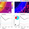
Samples of the non-magnetic fraction of a heavy mineral concentrate (HMC) concentration plant were analysed, as described by Potgieter-Vermaak.4 The samples originated specifically from the non-conducting fraction (Zr-rich) and were taken at various stages in its clean-up process. A sample that was supposed to be Ti-rich and practically free of Zr, was analysed by µRS, using the SEM-SCA instrument. Micro-Raman spectroscopic analyses showed that the Ti-rich part was comprised of both anatase and rutile. Some particles were covered in places with a red substance that appeared amorphous, potentially indicating secondary alteration; in some instances this was shown to be associated with anatase particles, but in other cases the fluorescence level was too high so that they could not be identified by µRS. As illustrated in Figure 4, the X-ray map indicated the co-existence of Al with anatase, which could not be identified by µRS. This maybe the reason for inefficient magnetic separation of these concentrates in the mineral industry.

Conclusions
SPA is an important tool in many scientific investigations. Not only can one determine the composition of particles from various sources to help answer questions about their role in atmospheric processes and events, one can also measure their main elemental compositions to establish whether they are inert or a potential threat to human health, ecosystems and the like. Interfacing elemental analyses with structural information provided by techniques such as µRS employed in conjunction with EPXMA, CC-SEM/EDX and XRF, further extends one’s ability to solve environmental, cultural heritage and even plant processing problems, and provides explanations for observed phenomena, as well as proposing solutions to them. The capability to probe both the elemental as well as the structural composition of materials on a micro-scale is essential to solve modern day research questions and provide answers about processes and mechanisms occurring on an atomistic scale. The examples discussed in this article serve to give the reader a small glimpse and taste of what can be accomplished by combining the analytical power of µRS with its corresponding EDX of single particles.
References
- L.A. De Bock, P.E. Joos, K.J. Noone, R.A. Pockalny and R.E. Van Grieken, “Single particle analysis of aerosols, observed in the marine boundary layer during the Moneterey Area Ship Tracks Experiment (MAST), with respect to cloud droplet formation”, J. Atmos. Environ. 37, 299–329 (2000).
- J. De Hoog, J. Osán, I. Szalóki, K. Eyckmans, A. Worobiec, C.-U. Ro and R. Van Grieken, “Thin-window electron probe X-ray micro-analysis of individual atmospheric particles above the North Sea”, Atmos. Environ. 39, 3231–3242 (2005).https://doi.org/10.1016/j.atmosenv.2005.02.025
- R.H.M. Godoi, S. Potgieter-Vermaak, J. De Hoog, R. Kaegi and R. Van Grieken, “Substrate selection for optimum qualitative and quantitative single atmospheric particle analysis, using nano-manipulation, sequential thin window electron probe micro-analysis and micro-Raman spectrometry”, Spectrochim. Acta B 61, 375–388 (2006).https://doi.org/10.1016/j.sab.2006.02.004
- S. Potgieter-Vermaak, “Surface characterization of a heavy mineral sand with micro-Raman spectrometry”, The 6th International Heavy Minerals Conference, “Back to Basics”. The Southern African Institute of Mining and Metallurgy, Marshalltown, South Africa (2007).