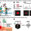
Gastrointestinal stromal tumours grow underneath the mucous layer covering our organs. Because they are deep inside the tissue, these “submucosal tumours” are difficult to detect and diagnose, even with a biopsy. Now, researchers from Japan have developed a novel minimally invasive and accurate method using infrared imaging and machine learning to distinguish between normal tissue and tumour areas. This technique has a strong potential for widespread clinical use.
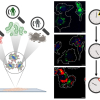
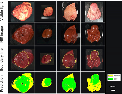
The team, led by Dr Hiroshi Takemura from Tokyo University of Science, performed imaging experiments on 12 patients with confirmed cases of GISTs, who had their tumours removed through surgery. The scientists imaged the excised tissues using NIR-HSI, and then had a pathologist examine the images to determine the border between normal and tumour tissue. These images were then used as training data for a machine-learning algorithm.
They found that even though 10 out of the 12 test tumours were completely or partly covered by a mucosal layer, the machine-learning analysis was effective in identifying GISTs, correctly classifying tumour and non-tumour sections with 86 % accuracy. “This is a very exciting development”, Dr Takemura explains, “being able to accurately, quickly and non-invasively diagnose different types of submucousal tumours without biopsies, a procedure that requires surgery, is much easier on both the patient and the physicians.”
Dr Takemura acknowledges that there are still challenges ahead, but feels they are prepared to solve them. The researchers identified several areas that would improve on their results, such as making their training dataset much larger, adding information about how deep the tumour is for the machine-learning algorithm, and including other types of tumours in the analysis. Work is also underway to develop an NIR-HSI system that builds on top of existing endoscopy technology.
“We’ve already built a device that attaches an NIR-HSI camera to the end of an endoscope and hope to perform NIR-HSI analysis directly on a patient soon, instead of just on tissues that had been surgically removed”, Dr Takemura says, “In the future, this will help us separate GISTs from other types of submucosal tumours that could be even more malignant and dangerous. This study is the first step towards much more ground-breaking research in the future, enabled by this interdisciplinary collaboration.”


