Electron paramagnetic resonance News
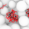
Scientists have succeeded for the first time in the direct spectroscopic detection of the binding of the “Parkinson protein” α-synuclein to lipid membranes in the cell.
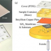
A new electron paramagnetic resonance (EPR) method only needs a very small liquid sample to provide detailed information on the state of the metal ions in a metalloprotein solution.
The 2017 Pittsburgh Spectroscopy Award has been made to Edward I. Solomon.
The protein α-synuclein plays an important role in Parkinson’s and other neurodegenerative diseases. Although a considerable amount is known about the structure of the protein within the Parkinson’s-typical amyloid deposits, nothing was known about its original state in the healthy cell up to now. High-resolution nuclear magnetic resonance and electron paramagnetic resonance spectroscopy have helped to visualise the protein in healthy cells.
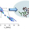
A new electron paramagnetic resonance (EPR) spectroscopy method is bringing researchers at Rensselaer Polytechnic Institute (RPI) closer to understanding—and artificially replicating—the solar water-splitting reaction at the heart of photosynthetic energy production.


